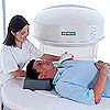
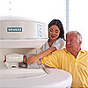
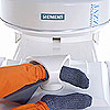
The New Magnetom Jazz is a patient-friendly orthopedic MR system from Siemens Medical Solutions
(MR patient positioning: shoulder, wrist, ankle and image montage: shoulder, ankle, elbow)
The New Magnetom Jazz is a patient-friendly orthopedic MR system from Siemens Medical Solutions
(MR patient positioning: shoulder, wrist, ankle and image montage: shoulder, ankle, elbow)
Magnetic Resonance (MR) imaging can non-invasively diagnose a number of sports related injuries without requiring surgery or arthroscopy. X-ray images of joints show good detail of bony structures and can clearly show a fracture. However, x-ray images give insufficient information about the soft tissue structures that hold joints together and allow the joint to support complex function. MR can yield detailed images and information on a number of bone and joint tissues like cartilage, tendon, ligament, bone marrow, bone cortex, muscle and joint fluids. With the development of new MR systems and techniques over the past five to ten years, MR has become an indispensable tool in diagnosing sports related injuries. MR imaging provides an optimal means to acquire high contrast images of both the bone structures and the soft tissue structures which together create a complex joint such as the knee or shoulder. More and more, MR imaging is being used as the main diagnostic tool in sports medicine while arthroscopy is being used as the guiding light for minimally invasive joint surgery.
Whole-body magnetic resonance imaging systems allow imaging of nearly every organ and structure in the human body. Whole body MR systems can image the brain, spine, liver, joints, extremities (limbs), blood vessels, heart and other organs in the body. However, these whole body systems are very expensive to own and operate because of their size and cost to manufacture. In the past few years, several medical systems manufacturers have developed new orthopedic MR systems that are primarily dedicated to imaging joints. This latest generation of extremity MR systems are very compact and combine orthopedic imaging performance with claustrophobia-free comfort for patients in a system that is far less expensive to own and operate. Further, these systems are small enough to be sited almost anywhere, even in the outpatient offices of orthopedic practices. Over the next several years these dedicated extremity systems will allow MR imaging to be conveniently accessible to more patients with orthopedic problems and sports related injuries. The image strip at the top of the page shows one of the newest orthopedic MR systems available.
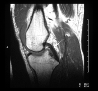
MR has incredible flexibility in its ability to selectively image and highlight specific tissues like the knee's meniscus or anterior cruciate ligament (ACL) and posterior cruciate ligament (PCL). MR has the unique ability to directly acquire images in almost any orientation so that surgeons can get a perfectly aligned view of a torn ACL. New three-dimensional MR (3D MR) techniques allow doctors to acquire an entire volume of data (for instance the entire knee). The doctors can then use the computer to go back and reconstruct images with the exact view or orientation that they need to see the pathology. "Musculoskeletal Imaging" is a specialized subset of MR imaging that is specifically geared towards imaging joints.
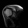
MR image of the knee showing posterior cruciate ligament (arrow)
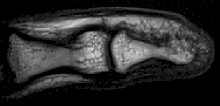
Musculoskeletal (or orthopedic) MR imaging is still a specialized field of MR and requires extensive experience and skill by the radiologist and technologist performing the MR exam. High quality musculoskeletal MR diagnosis also requires state-of-the-art MR systems, MR acquisition sequences and special so called "surface coils" which allow more detailed images of shoulder, knees, wrists and other complex joints. Even detailed MR images of the small joints and tendons in the finger can now be acquired.
High resolution MR image of the finger
Updated: November 2, 2007
This is health information, not medical advice.
Keep reading